
Lindenmann J, Matzi V, Neuboeck N, et al. Management of esophageal perforation in 120 consecutive patients: clinical impact of a structured treatment algorithm. J Gastrointest Surg. 2013 Jun;17(6):1036-43.
Full-text for Emory users.
 Click to enlarge.
Results: Iatrogenic perforation was the most frequent cause of esophageal perforation (58.3 %); Boerhaave’s syndrome was detected in 15 cases (6.8 %). Surgery was performed in 66 patients (55 %), 17 (14 %) patients received conservative treatment and 37 (31 %) patients underwent endoscopic stenting after tumorous perforation. Statistically significant impact on mean survival had Boerhaave’s syndrome (p = 0.005), initial sepsis (p = 0.002), pleural effusion/empyema (p = 0.001), mediastinitis (p = 0.003), peritonitis (p = 0.001), and redo-surgery (p = 0.000). Overall mortality rate was 11.7 %, in the esophagectomy group 17 % and in the patients with Boerhaave’s syndrome 33.3 %.
Conclusions: An approach considering etiology and extent of perforation, diagnostic delay, and septic status is required to improve patient’s outcome. Primary repair is feasible in patients without intrinsic esophageal disease and evidence of sepsis. The greater the diagnostic delay, the more the destruction of the esophageal wall especially in the case of septic esophageal disease, thus the stronger the argument for esophagectomy if anatomically and/or oncologically possible.
Kaman L, Iqbal J, Kundil B, Kochhar R. Management of Esophageal Perforation in Adults. Gastroenterology Res. 2010 Dec;3(6):235-244.
“Iatrogenic perforation is the leading cause of esophageal perforations [1–8] (Table 1). Iatrogenic causes account for around 70% of esophageal perforations [1, 3, 6, 8]. Endoscopic procedures are the most common cause of iatrogenic esophageal perforation. The reported risk for diagnostic esophagogastrodudenoscopy is 0.03%. The risk of perforation increases when therapeutic procedures are performed at the time of endoscopy. The reported risk for perforation is 0.5% in esophageal dilation, 1.7% in esophageal dilation for achalasia, 1-6% for endoscopic variceal sclerotherapy, 5% for endoscopic laser therapy, 4.6% for photodynamic therapy and esophageal stent placement carries a risk of 5-25% [1–8].”
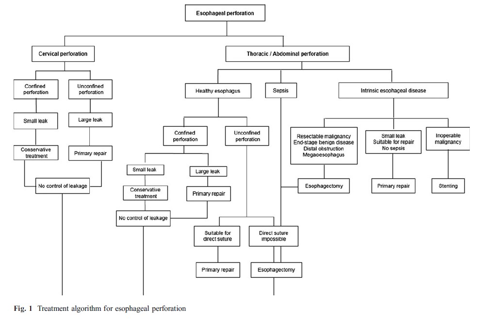
Figure 1. Management algorithm of esophageal perforation Click to enlarge.
