
Choledochal Cyst. In: Doherty GM. eds. Quick Answers Surgery. McGraw-Hill; Accessed April 02, 2021.
- Type I cysts (fusiform dilation of common bile duct [CBD]) account for 85-90%
- Type II (true diverticula of CBD) 1-2% of cases
- Type III (choledochocele—dilation of distal/intramural portion of CBD) < 2% of cases
- Type IV (multiple cysts involving intrahepatic and extrahepatic ducts) as high as 15% of cases in some series
- Type V (cystic malformation of intrahepatic ducts) rare
- 3-5% incidence of carcinoma
Soares KC, Arnaoutakis DJ, Kamel I, Rastegar N, Anders R, Maithel S, Pawlik TM. Choledochal cysts: presentation, clinical differentiation, and management. J Am Coll Surg. 2014 Dec;219(6):1167-80.
Full-text for Emory users.
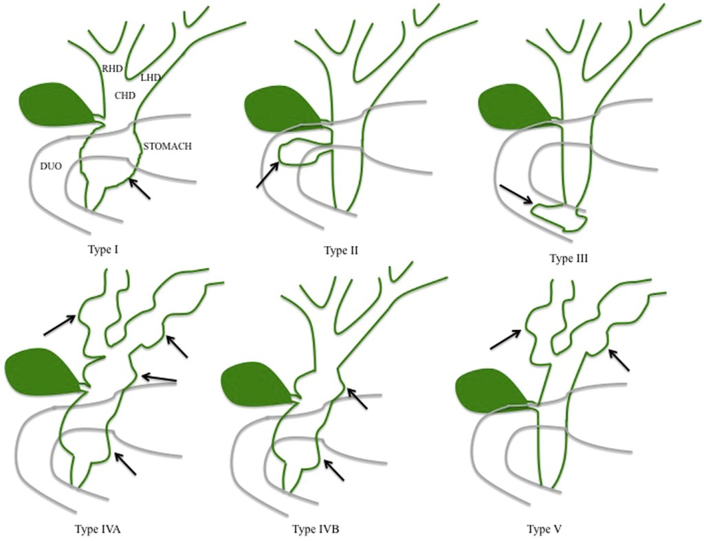
Type I cysts are fusiform dilatations of the common bile duct (CBD). Type II cysts are true diverticula of the CBD and type III CC (choledochoceles) are intraduodenal dilations of the common channel. Type IVA CC consist of multiple intrahepatic and extrahepatic biliary dilatations, while type IVB CC have extrahepatic biliary dilatation with a normal intrahepatic biliary tree. Type V CC, or Caroli’s disease, consist of cystic dilation of the intrahepatic biliary tree. RHD right hepatic duct, LHD left hepatic duct, CHD common hepatic duct, DUO duodenum.
See also: Clinical Overview: Choledochal cyst. Elsevier Point of Care. Updated October 7, 2019.
