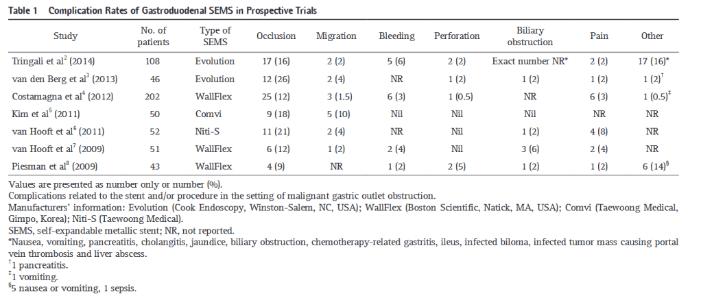
Covered stent placement for gastroduodenal artery stump hemorrhage after pancreaticoduodenectomy

“Post- pancreaticoduodenectomy (PD) hemorrhage is a rare but fatal complication that accounts for 10–40% of post-operative mortality.1,2 In such patients, successful surgical
treatment is compromised due to extensive inflammatory changes caused by recent dissections. Therefore, endovascular treatment is considered as the first line treatment
especially in cases of delayed hemorrhage (occurring 24 h after surgery) from the hepatic artery (HA). Transcatheter embolization and covered stent placement are the most
common endovascular techniques. However, transcatheter embolization typically involves sacrificing the major HA, which frequently causes severe hepatic ischemia or infarction.”

Continue reading

Emergency Colorectal Surgery in Those with Cirrhosis: Outcomes and predictors of mortality

“Individuals with cirrhosis have higher post-operative morbidity and mortality following major abdominal surgery compared to those without cirrhosis. To quantify this added risk, observational studies and prediction models have been described; however, the majority were derived from historic cohorts and may not reflect the changing epidemiology of
liver disease or advances in both the medical management of cirrhosis and perioperative practices in this high-risk group.”

Continue reading

Near‑infrared fluorescence cholangiography assisted laparoscopiccholecystectomy

“The most feared complication during laparoscopic cholecystectomy is bile duct injury. Bile duct injury as a result of laparoscopic cholecystectomy is rare with an incidence of 0.3–0.7% but often results in severe morbidity and even mortality, lower quality of life and extra costs.
Misidentification of extra-hepatic bile duct anatomy during laparoscopic cholecystectomy is the main cause of bile duct injury. Examples of such misidentification are mistaking the common bile duct for the cystic duct and aberrant hepatic ducts for the cystic duct or cystic artery. In order to reduce the risk of bile duct injury, techniques to enhance proper identification of the anatomy are needed.”

Continue reading

Comparative Safety of Robotic-Assisted vs Laparoscopic Cholecystectomy

“Whether robotic-assisted cholecystectomy leads to even safer outcomes than minimally invasive laparoscopic cholecystectomy remains unclear. Some contend that robotic-
assisted cholecystectomy may be safer because it offers 3-dimensional visualization, enhanced instrument articulation to allow for more complex maneuvers, novel ways to
visualize biliary anatomy, and potentially increases a surgeon’s ability to perform difficult procedures in a minimally invasive fashion. Studies comparing the safety of these
approaches found equivalency, but are limited to single-center case series inclusive of surgeons with the most robotic-assistance experience. Whether those outcomes reflect
current surgical practice, especially as robotic-assisted cholecystectomy is adopted by a larger and potentially more novice group of surgeons, represents crucial information for
surgeons, referring physicians, and patients.”

Continue reading

Robotic compared with laparoscopic cholecystectomy

“Robotic cholecystectomy was independently associated with a lower risk of serious complications, lower rate conversion to open, and hospitalization ≥24 hours compared with laparoscopic cholecystectomy. These findings suggest that new technologies might enhance the safety of minimally invasive surgery.”

Continue reading

Immediate versus Postponed Intervention for Infected Necrotizing Pancreatitis

“Acute pancreatitis is the most common pancreatic disease worldwide. Necrotizing pancreatitis develops in approximately 20 to 30% of patients with acute pancreatitis. Pancreatic and peripancreatic necrosis that becomes infected nearly always leads
to invasive intervention. The current standard approach for infected necrotizing pancreatitis is a minimally invasive step-up approach with catheter drainage as the first step. International guidelines advise postponement of catheter drainage and administration of antibiotics until the infected pancreatic and peripancreatic necrosis has become encapsulated; such walled-off necrosis usually takes 4 weeks to develop.”

Continue reading

Clinical characteristics and outcomes of patients with hepaticangiomyolipoma

“Angiomyolipoma (AML) is a solid mesenchymal tumor, mainly described in the
kidney, and belongs to the group of perivascular epithelioid cell tumors
(PEComas). Hepatic localization of AML, described for the first time in 1976, is
rare, since only around 600 cases were reported after an exhaustive search of the
literature up to the year 2017. Hepatic AML (HAML) poses a veritable diagnostic
challenge in radiological terms, especially when fat content is low, because this type of
tumor may appear as a hypervascular tumor associated with a washout phase that
mimics other, more common hypervascular hepatic tumors, such as hepatocellular
carcinoma”

Continue reading