Article of interest: The Landmark Series: MSLT-1, MSLT-2 and DeCOG (Management of Lymph Nodes)

Bello DM, Faries MB. The Landmark Series: MSLT-1, MSLT-2 and DeCOG (Management of Lymph Nodes). Ann Surg Oncol. 2020 Jan;27(1):15-21.

Full-text for Emory users.

Management of regional lymph nodes in patients with melanoma has evolved significantly in recent years. The value of nodal intervention, long utilized for its perceived therapeutic benefit, has now shifted to that of a critical prognostic procedure used to guide clinical decision making. This review focuses on the three landmark, randomized controlled trials evaluating the role of surgery for regional lymph nodes in melanoma: Multicenter Selective Lymphadenectomy Trial I (MSLT-I), German Dermatologic Cooperative Oncology Group-Selective Lymphadenectomy Trial (DeCOG-SLT), and Multicenter Selective Lymphadenectomy Trial II (MSLT-II).

Kikuchi-Fujimoto disease

Perry AM, Choi SM. Kikuchi-Fujimoto Disease: A Review. Arch Pathol Lab Med. 2018 Nov; 142(11):1341-1346.

Free full-text.

“Kikuchi-Fujimoto disease (KFD) is a rare entity characterized by subacute necrotizing lymphadenopathy and frequently associated with fever. Young adults of Asian ancestry are most commonly affected, but it has been reported worldwide. Despite many studies in the literature, the cause of KFD remains uncertain. Histologically, KFD is characterized by paracortical lymph node expansion with patchy, well-circumscribed areas of necrosis showing abundant karyorrhectic nuclear debris and absence of neutrophils and eosinophils. Three evolving histologic patterns — proliferative, necrotizing, and xanthomatous — have been recognized. By immunohistochemistry, histiocytes in KFD are positive for myeloperoxidase. There is a marked predominance of T cells in the lesions (with mostly CD8-positive cells) with very few B cells. The differential diagnosis of KFD includes infectious lymphadenitis, autoimmune lymphadenopathy (primarily systemic lupus erythematosus), and lymphoma.”

Continue reading

Articles of interest: Elimination of the Autopsy Requirement by CMS

Rueckert J. Elimination of the Autopsy Requirement by CMS. N Engl J Med. 2020 Feb 13;382(7):683-684.

Full-text for Emory users.

The autopsy plays a vital role in quality assurance by providing education and feedback to clinicians regarding diagnostic accuracy, therapeutic efficacy, and medical complications. At our institution, we promote a culture of transparency. Discrepant cases are discussed with the treating physicians and families and are also presented at conferences on morbidity and mortality, to educate a broad audience. As is the case at other academic institutions, our autopsy service provides extensive support of cutting-edge research efforts and hence is not “obsolete.”

CMS is making a mistake. A robust autopsy service plays an important role in providing and maintaining high-quality patient care. By eliminating the autopsy requirement, we are burying opportunities for improvement. The autopsy should be supported, not undermined.

Continue reading

NEJM Perspective: Learning from the Dead

De Cock KM, Zielinski-Gutiérrez E, Lucas SB. Learning from the Dead. N Engl J Med. 2019 Nov 14;381(20):1889-1891.

Full-text for Emory users.

“Obstacles to exploiting the rich database that the world’s decedents represent include not only the decline in autopsy rates but also a failure to prioritize broader innovative and culturally acceptable research and surveillance. Even information from medicolegal autopsies is not widely shared for auditing or educational purposes. A commitment among the medical and scientific communities to increase research and evaluation involving the dead, including assessments of postmortem investigations short of complete autopsies, could have great public health benefit.” (De Cock, 2019, pgs. 1889-1890)

Signet-ring cell carcinoma: a look at the rare colorectal cancer

A discussion this week included signet ring cell carcinoma.


Reference: Nitsche U, et al. Mucinous and signet-ring cell colorectal cancers differ from classical adenocarcinomas in tumor biology and prognosis. Annals of Surgery. 2013 Nov;258(5):775-782; discussion 782-783. doi:10.1097/SLA.0b013e3182a69f7e

Additional Reading: Korphaisarn K, et al. Signet ring cell colorectal cancer: genomic insights into a rare subpopulation of colorectal adenocarcinoma. British Journal of Cancer. 2019 Sep;121(6):505-510. doi:10.1038/s41416-019-0548-9

Summary: In a study analyzing clinical, histopathological, and survival data of 3479 patients undergoing surgery for primary colorectal cancer between 1982 and 2012, Nitsche et al (2013) compared the characteristics of classical adenocarcinomas (AC) to the less common mucinous adenocarcinomas (MAC) and to the rare signet-ring cell carcinomas (SC).

SC

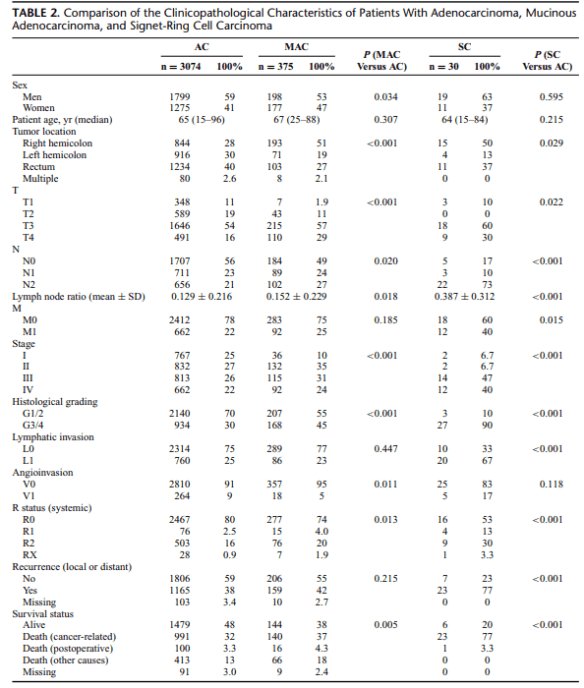
Approximately 10% of all colorectal cancers are MAC, and about 1% are SC. Because of their relatively rare occurrence, in particular, the evaluation of the clinical impact of SC is difficult. However, compared with AC, both MAC and SC have been shown to be associated with young age, advanced tumor stage, accumulation in female patients, and distinct molecular patterns, such as microsatellite instability and activating mutations of the BRAF gene. Although ambiguous, recent data and meta-analyses suggest that the
histological subtype MAC may be associated with worse outcome compared with AC. Poor prognosis of SC is more evident, mainly due to high rates of synchronous and metachronous distant organ metastasis associated with this histological subtype.

In describing SC, the authors state: “SC have been described as being positive for intestinal trefoil factor and MUC2, 2 peptides that are usually produced only by goblet cells. Thus, SC could arise from different cells of origin than AC. Although they can be localized in the colorectum, SC may be genetically more related to signet-ring cell cancers of other organs (eg, gastric cancer) than to AC or MAC of the colorectum. The
absence of E-cadherin/β-catenin and amplification of Bcl-2 are features typically shared with signet-ring cell cancer of the stomach but not with classical colorectal adenocarcinomas” (p.781).

The authors conclude that patients with MAC and SC could profit from closer follow-up or even intensified adjuvant therapy because of their high rates of local and distant recurrence. The biological behavior of SC differs in specific, and these patients require special awareness, despite the relatively rare prevalence.

Air cholangiogram as effective measure for postoperative biliary complications

One discussion this week involved air cholangiograms.


Reference: Zimmitti G, et al. Systematic use of an intraoperative air leak test at the time of major liver resection reduces the rate of postoperative biliary complications. Journal of the American College of Surgeons. 2013 Dec;217(6):1028-1037. doi: 10.1016/j.jamcollsurg.2013.07.392.

Summary: Advances in surgical technique and better understanding of liver anatomy and physiology have facilitated a decrease in postoperative hepatic insufficiency rates and in perioperative blood transfusion needs. However, these improvements have not been paralleled by a decrease in the rate of postoperative bile leak, which remains the Achilles’ heel of liver resection. While in many cases a postoperative bile leak can be managed successfully with drainage and antibiotics, it almost always entails longer length of stay and increased hospital costs.

Continue reading

A review of ICPN (by Emory pathologists!)

One discussion this week included ICPN.

Reference: Adsay V, et al. Intracholecystic papillary-tubular neoplasms (ICPN) of the gallbladder (neoplastic polyps, adenomas, and papillary neoplasms that are ≥1.0 cm): clinicopathic and immunohistochemical analysis of 123 cases. The American Journal of Surgical Pathology. 2012 Sep;36(9):1279-1301.

Summary (from the abstract):  In this study, 123 GB cases that have a well-defined exophytic preinvasive neoplasm measuring ≥1 cm were analyzed. The patients were predominantly female (F/M=2:1) with a mean age of 61 y and a median tumor size of 2.2 cm. Half of the patients presented with pain, and in the other half the neoplasm was detected incidentally. Other neoplasms, most being gastrointestinal tract malignancies, were present in 22% of cases. Gallstones were identified in only 20% of cases. Radiologically, almost half were diagnosed as “cancer,” roughly half with polypoid tumor, and in 10% the lesion was missed.

Pathologic Findings 

  1. The predominant configuration was papillary in 43%, tubulopapillary in 31%, tubular in 26%.
  2. Each case was assigned a final lineage type on the basis of the predominant pattern (>75% of the lesion) on morphology, and supported with specific immunohistochemical cell lineage markers. The predominant cell lineage could be identified as biliary in 50% (66% of which were MUC1), gastric foveolar in 16% (all were MUC5AC), gastric pyloric in 20% (92% MUC6), intestinal in 8% (100% CK20; 75% CDX2; 50%, MUC2), and oncocytic in 6% (17% HepPar and 17% MUC6); however, 90% of cases had some amount of secondary or unclassifiable pattern and hybrid immunophenotypes.
  3. Of the cases that would have qualified as “pyloric gland adenoma,” 21/24 (88%) had at least focal high-grade dysplasia and 18% had associated invasive carcinoma. Conversely, 8 of 47 “papillary adenocarcinoma”-type cases displayed some foci of low-grade dysplasia, and 15/47 (32%) had no identifiable invasion.
  4. Overall, 55% of the cases had an associated invasive carcinoma (pancreatobiliary type, 58; others, 10). Factors associated significantly with invasion were the extent of high-grade dysplasia, cell type (biliary or foveolar), and papilla formation. Among systematically analyzed invasive carcinomas, tumoral intraepithelial neoplasia was detected in 6.4% (39/606).
  5. The 3-year actuarial survival was 90% for cases without invasion and 60% for those associated with invasion. In contrast, those associated with invasion had a far better clinical outcome compared with pancreatobiliary-type GB carcinomas (3-yr survival, 27%), and this survival advantage persisted even with stage-matched comparison. Death occurred in long-term follow-up even in a few noninvasive cases (4/55; median 73.5 mo) emphasizing the importance of long-term follow-up.

Conclusion

Tumoral preinvasive neoplasms (≥1 cm) in the GB are analogous to their pancreatic and biliary counterparts (biliary intraductal papillary neoplasms, pancreatic intraductal papillary mucinous neoplasms, and intraductal tubulopapillary neoplasms). They show variable cellular lineages, a spectrum of dysplasia, and a mixture of papillary or tubular growth patterns, often with significant overlap, warranting their classification under 1 unified parallel category, intracholecystic papillary-tubular neoplasm. Intracholecystic papillary-tubular neoplasms are relatively indolent neoplasia with significantly better prognosis compared with pancreatobiliary-type GB carcinomas. In contrast, even seemingly innocuous examples such as those referred to as “pyloric gland adenomas” can progress to carcinoma and be associated with invasion and fatal outcome.