SAGES guidelines for the use of intraoperative imaging of the common bile duct

“The purpose of these guidelines is to provide evidence-based recommendations for the use of intraoperative imaging modalities during laparoscopic cholecystectomy. We assessed surgical outcomes that were deemed to be important to both surgeons and patients by a panel of surgeons. Specifically, the imaging modalities investigated were contrast
intraoperative cholangiography (IOC), fluorescence imaging (FI) with indocyanine green (ICG), and laparoscopic ultrasound (LUS). The target audience for these guidelines
includes surgeons and patients. A patient–physician perspective was taken, so cost-effectiveness, resources requirements, and availability of said resources were not evaluated. These imaging modalities were primarily assessed in the context of the American healthcare system. Therefore, these recommendations may not apply in settings where the relevant
technology is not easily accessible.”

“Laparoscopic cholecystectomy is one of the most common abdominal operations performed in the United States. Injury to the common bile duct is a rare but dreaded complication, often requiring additional surgery or endoscopic intervention. Its occurrence has been increasing since the introduction and widespread adoption of laparoscopy
(0.4–1.5% of cases) compared to open cholecystectomy. Multiple factors may contribute to the risk of common bile duct injury, including the presence of acute inflammation and anatomical variability of the CBD. Strategies such as achieving the Critical View of Safety (CVS) were developed to mitigate this risk.”
Continue reading

Routine or Selective Intraoperative Cholangiography?

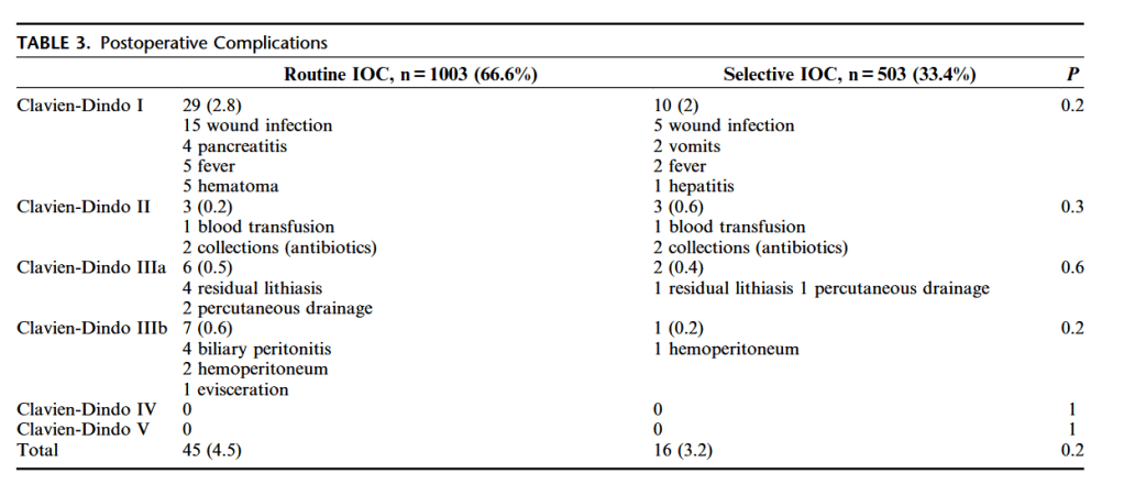
“Intraoperative cholangiography (IOC) helps to clarify the biliary tree anatomy, detect common bile duct (CBD) stones, and may prevent or promptly detect BDI. However,
the use of routine (RC) or selective (SC) IOC remains controversial. Some authors have suggested that RC allows for early detection of BDI and repair in the same surgical
procedure, reducing morbidity and mortality.6–8 Also, IOC facilitates the detection and treatment of CBD stones, reducing potential complications and readmissions. In
contrast, opponents emphasize that RC often detects asymptomatic CBD stones leading to unnecessary procedures, increases operating time, costs, and exposes the surgical team and patients to radiation.”

Continue reading

Advantages of routine intraoperative cholangiography in a teaching hospital

“The role of routine IOC during cholecystectomy has been controversial. Opponents to routine IOC assert that this procedure increases operating times and exposes caregivers and patients to radiation. In addition, there is the possibility of detection of indolent CBD stones with consequently unnecessary removal. On the other hand, advocates in favor
of routine IOC state that intraoperative visualization of the bile duct anatomy may decrease either the rate of complications such as CBD injury, or hospital readmissions for subsequent removal of retained CBD stones. Despite lacking strong evidence for not performing IOC vs. routine IOC vs. selective IOC, fitting in one of these three groups can depend on training, technical experience, and surgical habit. If a surgeon never performs IOC in their daily practice, they are not eager to change their habits, even though literature may suggest otherwise.”

Continue reading

Near‑infrared fluorescence cholangiography assisted laparoscopiccholecystectomy

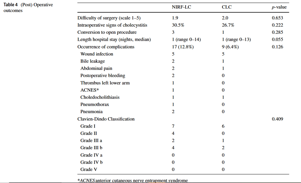
“The most feared complication during laparoscopic cholecystectomy is bile duct injury. Bile duct injury as a result of laparoscopic cholecystectomy is rare with an incidence of 0.3–0.7% but often results in severe morbidity and even mortality, lower quality of life and extra costs.
Misidentification of extra-hepatic bile duct anatomy during laparoscopic cholecystectomy is the main cause of bile duct injury. Examples of such misidentification are mistaking the common bile duct for the cystic duct and aberrant hepatic ducts for the cystic duct or cystic artery. In order to reduce the risk of bile duct injury, techniques to enhance proper identification of the anatomy are needed.”

Continue reading

Intraoperative cholangiography during laparoscopiccholecystectomy:

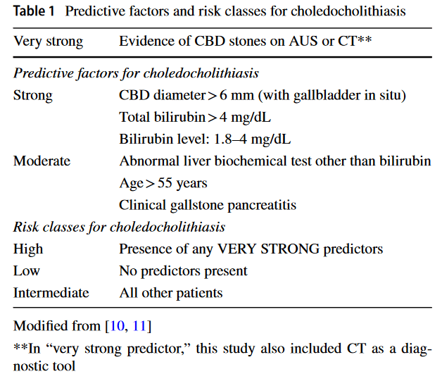
“Based on the study results, the 2016 WSES risk classes for choledocholithiasis could be an effective approach for predicting the risk of choledocholithiasis. Considering its advantages for detecting CBD stones and biliary injuries, the routine use of IOC is still suggested.” (Lai)

(Lai)
Continue reading

Incidence of problematic common bile duct calculi in patients undergoing laparoscopic cholecystectomy.

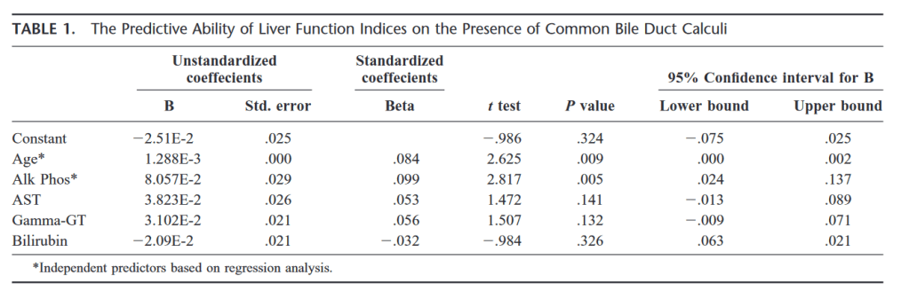
“Choledocholithiasis occurs in 3.4% of patients undergoing laparoscopic cholecystectomy but more than one third of these pass the calculi spontaneously within 6 weeks of operation and may be spared endoscopic retrograde cholangiopancreatography.” (Collins)

(Collins)
Continue reading

What is the utility of routine intraoperative cholangiography during laparoscopic cholecystectomy?

SAGES still recommends that practicing general surgeons learn how to do IOC (though once a surgeon is past their learning curve, it is not necessarily routinely recommended that it be done ‘routinely’).


Hope WW, et al. SAGES clinical spotlight review: intraoperative cholangiography. Surg Endosc. 2017 May;31(5): 2007-2016. Full-text for Emory users.

“The following clinical spotlight review regarding the intraoperative cholangiogram is intended for physicians who manage and treat gallbladder/biliary pathology and perform laparoscopic cholecystectomy. It is meant to critically review the technique of intraoperative cholangiography, alternatives for intraoperative biliary imaging, and the available evidence supporting their safety and efficacy.”

Continue reading