What is the operative management of diverticulitis?

One discussion this week included the operative management of diverticulitis.

Reference: Nally DM and Kavanagh DO. Current controversies in the management of diverticulitis: a review. Digestive Surgery. 2018 Apr 19. doi:10.1159/000488216.

Summary: A treatment algorithm is illustrated below (Nally and Kavanagh, 2018, p.7).

fig2

Laparoscopic Lavage

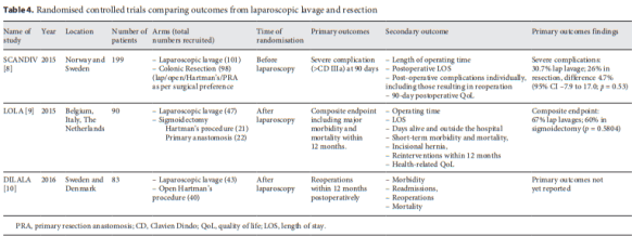
Nally and Kavanagh highlight three RCTs comparing laparoscopic lavage and resection (Table 4, p.8): SCANDIV (2015), LOLA (2015), and DILALA (2016).  The main differences in the RCTs were the re-intervention rate, which varied according to when results were reports (prior to the time of expected stoma closure) or if stoma closure is specifically excluded. Surgical reinterventions accounted for a majority of adverse events.

table4

Surgical Resection

Originally a 3-stage operation, it was condensed into a 2-stage Harman’s procuedure in which the diseased segment was removed during initial laparotomy. This became the standard of care, yet the mortality rate is cited as 15% according to the SCANDIV study. Additionally, a Hartman’s procedure is limited by the morbidity and mortality of colostomy reversal or lifestyle implications of a permanent stoma for up to 60% of patients (p.8). A one-stage resection with restoration of continuity avoids some challenges but also creates concern about performing an anastomosis in a contaminated environment for a critically unwell patient.

A 2004 systematic review found an overall mortality rate of 9.9% for primary anastomosis  (n=568) and 18.8% for Hartman’s (n=1,051), with overall anastomotic leak rate for a primary anastomosis of 13.9% (p.8).

Elective Surgery for Diverticulitis

Approximately 15-30% of patients have recurrent episodes of diverticulities. Up to 30% have ongoing pain. There are 2 indications for elective surgery:

  1. Prophylaxis against recurrent attacks and complications
  2. Surgery for ongoing symptoms that impact quality of life.

Guidelines from the American Society of Surgeons of Colon and Rectal surgeons from 2000 recommended an elective resection after one or two episodes of acute uncomplicated diverticulitis (p.9). Currently, professional bodies included the AGA advise again routine resection and instead propose an individualized approach (p.9). Morbidity and mortality of elective procedures for diverticulitis can be significant.

Conversely, elective surgery to improve quality of life is coming more to the fore (p.9). A systematic review of 21 studies and more than 1,800 patients and an RCT – the DIRECT trial – with 109 patients seem to provide evidence in favor of resection. It must be noted that the RCT was terminated early.

Nally and Kavanagh (2018) conclude that diverticulitis is a common yet challenging topic that demands clinicians to provide an individualized yet evidence-based approach (p.1).

The use of REBOA for trauma

One discussion this week included the use of REBOA for trauma cases.

Reference: Brenner, M, et al. Use of resuscitative endovascular balloon occlusion of the aorta for proximal aortic control in patients with severe hemorrhage and arrest. JAMA Surgery. 2018 Feb;153(2):130-135. doi:10.1001/jamasurg.2017.3549.

Summary: Reporting on the largest single-institution study on REBOA in the US, Brenner et al (2018) state that the risks of clinician exposure and morbidity of opening the thorax to cross-clamp the aorta make REBOA a more attractive option than emergency department thoracotomy with aortic cross-clamp (EDTCC). This study observed outcomes of patients with  severe traumatic hemorrhage, traumatic arrest (AR), and nontraumatic hemorrhage (NTH) between 2013 and 2017.

For 79 patients with severe traumatic hemorrhage and AR, in-hospital mortality was 71%. Technical success, as defined by AO at the intended level (zone 1 or 3), occurred in 44 of the 53 patients (83%) who had radiographic, fluoroscopic, manual, or CT confirmation of the balloon. The remaining identified malpositioned catheters were repositioned immediately to a slightly more proximal location (proximal zone 2 to distal zone 1) without clinical sequelae. Seven patients underwent REBOA at zone 1, which was then purposefully repositioned to zone 3 after intra-abdominal hemorrhage was ruled out by imaging (n = 3) or surgical exploration (n = 4) (p.131-132).

In the patients with severe traumatic hemorrhage, the 30-day survival was 59% (p.132). Indications for REBOA were transient responders or nonresponders who remained severely hypotensive despite resuscitation efforts. A total of 18 patients (62%) received REBOA in zone 1, while 11 patients with severe hemorrhage from the pelvis or below (38%) received REBOA in zone 3. Twelve patients received REBOA in the OR; the indications included AR or impending AR, refractory hypotension, presence of expanding pelvic hematoma with abdominal hemostasis, and performance of REBOA prior to exploration of a large central hematoma including, in 1 patient, severe adhesions from a previous laparotomy.

Of the patients with AR, 50 received REBOA while in arrest. Spontaneous circulation occurred in 29 (58%), 20 of those survived to the OR. The 30-day survival was 10% (p.133). Access to the CFA was percutaneous in 13 patients and via surgical cutdown
in 37 patients, including 8 patients who had access attempted percutaneously but completed via cutdown. Patients received cardiopulmonary resuscitation throughout the REBOA procedure (p.133).

Benefits of REBOA are:

  • the ability to provide continuous closed chest compressions during the procedure
  • its ability to temporize hemorrhage and thus buy time to gather results of diagnostic imaging, especially when other injuries may alter treatment algorithms
  • the consequences of extended occlusion, particularly in patient care settings without resources for definitive hemorrhage control

Brenner et al (2018) note that REBOA can also be used for more targeted AO in the distal aorta for pelvic, junctional, or extremity hemorrhage (p.135).

What is the morbidity and mortality of TEVAR for ruptured thoracic aortic aneurysms?

One discussion this week included the morbidity and mortality of TEVAR in the setting of ruptured thoracic aortic aneurysms.

Reference: Geisbusch, P, et al. Endovascular repair of ruptured thoracic aortic aneurysms is associated with high perioperative mortality and morbidity. Journal of Vascular Surgery. 2010 Feb;51(2):299-304. doi:10.1016/j.jvs.2009.08.049.

Summary: In a retrospective study, Geisbusch et al (2010) analyzed the outcomes of emergency endovascular treatment of thoracic aortic pathologies (TEVAR). Out of 236 patients, 23 received thoracic aortic repair due to a ruptured thoracic aortic aneurysm (rTAA). The overall hospital mortality was 48% (see table below, p.302). Overall technical success was 87%. Three patients showed relevant primary endoleaks, and thus were not considered technical successes.

table3

The causes of death in the 11 patients were: cardiac complications (7), multiorgan failure (3), and pulmonary embolism (1) (p.301).

The authors admit: “Mortality rates after TEVAR for acute descending aortic rupture vary between 0% and 17% in the few available series, which seems relatively low compared with our in-hospital mortality rate of 48%” (p.303).

The patient population in this study had a median age of 75 years, and was highly comorbid (83% with coronary heart disease, 43% with renal insufficiency, and 30% with COPD), resulting in high cardio-pulmonary and renal complications with consecutive perioperative death. Three-year survival is estimated at 30%.

In conclusion, “the endovascular treatment of ruptured thoracic aortic aneurysms is associated with a high perioperative mortality and morbidity as well as poor midterm survival. Renal insufficiency proved as an independent risk factor for perioperative death” (p.303).

Open abdomen: indications and management

One discussion this week included open abdomen (OA) management.

Reference: Coccolini F, et al. The open abdomen in trauma and non-trauma patients: WSES guidelines. World Journal of Emergency Surgery. 2018 Feb 2;13:7. doi:10.1186/s13017-018-0167-4.

Summary: The table below summarizes the guidelines statements, including grades of evidence. Please note, these guidelines are methods for optimal management and are not a standard of practice (p.2).

Indications (p.4):

table2 indications

Closure (p.5):

Table2

Sclerosing encapsulating peritonitis

One discussion this week involved small bowel obstruction and sclerosing encapsulating peritonitis (SEP).


Reference: Liberale G, Sugarbaker PH. Sclerosing encapsulating peritonitis as a potential complication of cytoreductive surgery and HIPEC: clinical features and results of treatment in 4 patients. Surgical Oncology. 2018 Dec;27(4):657-662.

Summary: Liberale and Sugarbaker (2018) define SEP as “a rare entity characterized by encapsulation of the small bowel and/or the colon by fibrous tissue forming a shell” (p.657). It is iatrogenic, idiopathic, or secondardy, and its pathophysiology is uncertain. The most common symptoms are abdominal pain, discomfort, and partial or complete obstruction.

In this article, the authors present 4 cases of SEP, all of which required additional surgery to alleviate recurrent episodes of small bowel obstruction.

In discussion, they provide some advice (p.661):

  • An adverse event to avoid is small bowel fistula following surgery.
  • The prevention of fistulization which results in enteric contamination of the peritoneal space is of utmost importance in reoperative surgery.
  • Careful marking of seromuscular tears and their repair prior to closing the abdomen is important.
  • A major problem that may occur in follow-up is the difficulty of distinguishing recurrence of peritoneal metastases from benign causes of bowel obstruction.

There are two types of SEP (p.661):

  • Type I: a fibrous membrane sheathing the bowel loops together without a clearly separated dissection plane. Surgery is challenging and the surgeon needs to open the plane between bowel loops while avoiding causing serosal tears.
  • Type II: a fibrous membrane forming an enterocele or ‘pseudocyst-like’ structure. These are easier to manage as, once the pouch is open, the small bowel can be dissected and separated easily from the surrounding sheath.

The Carotid Revascularization Endarterectomy vs Stenting Trial (CREST)

One discussion this week involved the Carotid Revascularization Endarterectomy vs. Stenting Trial (CREST).


Reference: Brott TG, et al. Stenting versus endarterectomy for treatment of carotid-artery stenosis. New England Journal of Medicine. 2010 Jul 1;363(1):11-23. doi:10.1056/NEJMoa0912321.

Summary:  CREST is an RCT with blinded end-point adjudication whose aim was “to compare the outcomes of carotid-artery stenting with those of carotid endarterectomy among patients with symptomatic or asymptomatic extracranial carotid stenosis” (p.12).

Between December 2000 through July 2008, 2522 patients were enrolled in 108 centers in the US and 9 in Canada. Of those, 1271 patients were randomly assigned to undergo carotid-artery stenting.

Primary findings include (p.18):

  • Carotid revascularization performed by highly qualified surgeons and interventionists is effective and safe.
  • Stroke was more likely after carotid-artery stenting.
  • Myocardial infarction was more likely after carotid endarterectomy, but the effect on the quality of life was less than the effect of stroke.
  • Younger patients had slightly fewer events after carotid-artery stenting than after carotid endarterectomy.
  • Older patients had few events after carotid endarterectomy.
  • Low absolute risk of recurrent stroke suggests that both carotid-artery stenting and carotid endarterectomy are clinically durable and reflect advances in medical therapy.