Evaluating the Role of Indocyanine Green Fluorescence Imaging in Enhancing Safety and Efficacy During Laparoscopic Cholecystectomy

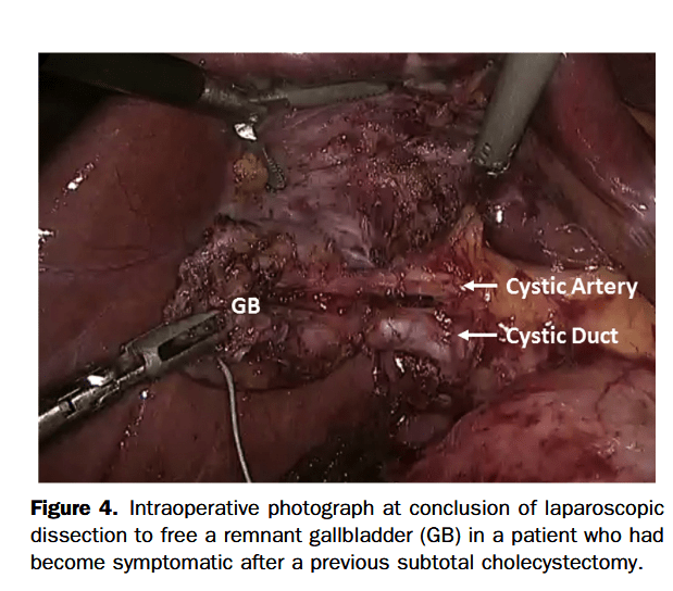
“In recent years, indocyanine green (ICG) fluorescence imaging has emerged as an alternative tool to enhance the visualization of biliary structures during LC. ICG is a fluorescent dye that, when injected intravenously, is preferentially taken up by the liver and excreted into the bile ducts. When exposed to near-infrared light, ICG causes the biliary structures, such as the CD, CBD, and CA, to fluoresce, making them more distinguishable from surrounding tissues thereby facilitating real-time visualization of biliary structures during the dissection of Calot’s triangle. The timing of ICG injection is critical to ensure that the biliary anatomy lights up distinctly without interference from non-biliary structures.
However, the routine use of ICG fluorescence imaging in LC has not yet been standardized, and there is ongoing debate about whether its widespread adoption would significantly reduce the incidence of BDI and improve patient outcomes. This systematic review aims to provide a comprehensive evaluation of the efficacy and safety of ICG fluorescence imaging in LC, specifically comparing its impact on the incidence of BDI to that of conventional white light (WL) imaging.”

Table 4. Comparison of visualization of biliary structures and incidence of BDI using ICG fluorescence vs WL in LC.

BDI, bile duct injury; CBD, common bile duct; CD, cystic duct; CHD, common hepatic duct; ICG, indocyanine green; LC, laparoscopic cholecystectomy; WL, white light; -, not specified

Author(s) and yearVisualization of CDVisualization of CBDVisualization of CHDVisualization of the CD-CBD junctionIncidence of BDI using ICGIncidence of BDI using WL
Symeonidis et al., 2024No significant difference (p = 0.225)No significant difference (p = 0.276)No significant difference (p = 0.940)No significant difference (p = 0.827)00
Ma et al., 2023 Before dissecting Calot’s: no significant difference (p = 0.075). After dissecting Calot’s: ICG signifi-cantly improved visualization (p = 0.02)Before dissecting Calot’s: no significant difference (p = 0.075). After dissecting Calot’s: ICG signifi-cantly improved visualization (p = 0.02)00
Xu et al., 2023 00
Stolz et al., 2023 No significant differenceNo significant differenceNo significant differenceNo significant difference
Lie et al., 2023 Improved RR 1.24, 95% CI 1.07–1.43, p = 0.003Improved: RR 1.31, 95% CI 1.07–1.60, p = 0.009No significant difference: (RR 0.34, 95% CI 0.07–1.58, p = 0.17)  No significant difference: (RR 0.34, 95% CI 0.07–1.58, p = 0.17)
Losurdo et al., 2022 01.4%, p = 0.728
Lacuzzo et al., 2022 00
Jin et al., 2022 01.83%, p = 0.389
Lim et al., 2021No significant difference: RR = 0.90, p = 0.12, 95% CI 0.79– 1.03, I² = 74%No significant difference: RR = 0.82, p = 0.09, 95% CI 0.65– 1.03, I² = 87%ICG significantly improved visualization: RR = 0.58, p = 0.03, 95% CI 0.35–0.93, I² = 91%No significant difference: RR = 0.68, p = 0.06, 95% CI 0.45– 1.02, I² = 94%02 (0.55%)
Dip et al., 2021 1 (0.06%)12 (0.25%)
Broderick et al., 2021 01 (0.1%), p = 1  
Keeratibharat, 2021 ICG signifi-cantly improved visualization, p = 0.001ICG signifi-cantly improved visualization, p = 0.002ICG signifi-cantly improved visualization, p = 0.00000
Ambe et al., 2019 00
Dip et al., 2019 Before dissecting Calot’s: ICG signifi-cantly improved visualization (p ≤ 0.001). After dissecting Calot’s: no significant difference (p = 0.83)Before and after dissecting Calot’s: ICG signifi-cantly improved visualization (p < 0.001)Before and after dissecting Calot’s: ICG signifi-cantly improved visualization (p < 0.001)Before and after dissecting Calot’s: ICG signifi-cantly improved visualization (p < 0.001)02 (0.62%)
Continue reading

Subtotal Cholecystectomye“Fenestrating” vs“Reconstituting” Subtypes and the Prevention ofBile Duct Injury

“Laparoscopic cholecystectomy is a well-established procedure with clear benefits for patients over open cholecystectomy. However, it is associated with an increased rate of
bile duct injury. Biliary injuries occur more commonly when operations are made more difficult due to the presence of severe acute and/or chronic inflammation. Under these conditions, secure ductal identification by the critical view of safety (CVS) may be very challenging because CVS requires clearing of the inflamed hepatocystic triangle in
order to demonstrate the cystic duct, cystic artery, and the cystic plate. It is a rigorous method, but as we have previously stressed, this is actually one of the strengths of the
CVS method of identification. The infundibular technique, in which the funnel-shaped infundibular-cystic duct junction is the rationale for identification, is much easier to achieve than CVS.”

Continue reading

Patient Complexity and Bile Duct Injury After Robotic-Assisted vs Laparoscopic Cholecystectomy

“Whether robotic-assisted cholecystectomy offers an advantage over laparoscopic cholecystectomy for higher-risk cases remains unclear. On one hand, there may be fundamental differences in the complexity of patients undergoing robotic-assisted cholecystectomy, which may be responsible for the higher observed rates of bile duct injury. On the other hand, differences in bile duct injury could be secondary to other factors, such as surgeons working their way up the learning curve using the robot, especially given the large number of robotic-assisted cholecystectomies surgeons must perform to achieve bile duct injury rates equivalent to those of laparoscopic approaches. By comparing laparoscopic and robotic-assisted cholecystectomy approaches within patient risk terciles, we can determine whether patient risk factor profiles are associated with harm in robotic-assisted cholecystectomy.”

Continue reading

Near‑infrared fluorescence cholangiography assisted laparoscopiccholecystectomy

“The most feared complication during laparoscopic cholecystectomy is bile duct injury. Bile duct injury as a result of laparoscopic cholecystectomy is rare with an incidence of 0.3–0.7% but often results in severe morbidity and even mortality, lower quality of life and extra costs.
Misidentification of extra-hepatic bile duct anatomy during laparoscopic cholecystectomy is the main cause of bile duct injury. Examples of such misidentification are mistaking the common bile duct for the cystic duct and aberrant hepatic ducts for the cystic duct or cystic artery. In order to reduce the risk of bile duct injury, techniques to enhance proper identification of the anatomy are needed.”

Continue reading

Comparative Safety of Robotic-Assisted vs Laparoscopic Cholecystectomy

“Whether robotic-assisted cholecystectomy leads to even safer outcomes than minimally invasive laparoscopic cholecystectomy remains unclear. Some contend that robotic-
assisted cholecystectomy may be safer because it offers 3-dimensional visualization, enhanced instrument articulation to allow for more complex maneuvers, novel ways to
visualize biliary anatomy, and potentially increases a surgeon’s ability to perform difficult procedures in a minimally invasive fashion. Studies comparing the safety of these
approaches found equivalency, but are limited to single-center case series inclusive of surgeons with the most robotic-assistance experience. Whether those outcomes reflect
current surgical practice, especially as robotic-assisted cholecystectomy is adopted by a larger and potentially more novice group of surgeons, represents crucial information for
surgeons, referring physicians, and patients.”

Continue reading

Robotic compared with laparoscopic cholecystectomy

“Robotic cholecystectomy was independently associated with a lower risk of serious complications, lower rate conversion to open, and hospitalization ≥24 hours compared with laparoscopic cholecystectomy. These findings suggest that new technologies might enhance the safety of minimally invasive surgery.”

Continue reading

Laparoscopic Entry Techniques and Injuries

“Recent reports by the Australian Safety and Efficacy Register for New interventions and Procedures (ASERNIP-S) and the Royal Australian and New Zealand College of Obstetricians and Gynaecologists (RANZCOG) concluded that insufficient evidence is available to assess the safety of the open versus closed laparoscopy in regard to major vascular and visceral injuries.” (Larobina & Nottle)

Major Vascular Injuries in Closed vs. Open Laparoscopy (Larobina & Nottle)

“Our case series shows that open laparoscopy can vastly reduce the incidence of access-related morbidity and mortality. Only a single visceral injury occurred in 5900 cases, and no major vascular injuries were reported.These figures are consistent with those of other reported series of open laparoscopy, which also show a zero rate of vascular injury and low rates of visceral injury.The literature review showed a rate of 1 injury to major retroperitoneal vessels per 2272 cases of closed laparoscopy procedures. This compares with a major vascular injury rate of 0 for the open technique. This difference is both statistically significant ( P = 0.003) and highly clinically significant.” (Larobina & Nottle)

Continue reading