Stomal necrosis

Murken DR, Bleier JIS. Ostomy-Related Complications. Clin Colon Rectal Surg. 2019 May;32(3):176-182.

Full-text for Emory users.

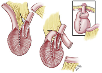
“Stomal necrosis has been reported to occur in up to 20% of ostomates in the immediate postoperative period ([Fig. 1]).[3] Specific risk factors for stoma necrosis include emergent operation, inadequate mobilization of the bowel, excessive mesenteric resection resulting in inadequate arterial blood supply to or venous drainage from the bowel, and constriction in the abdominal wall due to excessively small openings in the fascia, abdominal wall mesh, or skin.[10] [20] Importantly, the obese patient is seven times more likely to experience stoma necrosis than the nonobese patient.[21] Stoma necrosis is much less common for loop stomas given the dual blood supply to both the afferent and efferent limbs.”

Continue reading

Kikuchi-Fujimoto disease

Perry AM, Choi SM. Kikuchi-Fujimoto Disease: A Review. Arch Pathol Lab Med. 2018 Nov; 142(11):1341-1346.

Free full-text.

“Kikuchi-Fujimoto disease (KFD) is a rare entity characterized by subacute necrotizing lymphadenopathy and frequently associated with fever. Young adults of Asian ancestry are most commonly affected, but it has been reported worldwide. Despite many studies in the literature, the cause of KFD remains uncertain. Histologically, KFD is characterized by paracortical lymph node expansion with patchy, well-circumscribed areas of necrosis showing abundant karyorrhectic nuclear debris and absence of neutrophils and eosinophils. Three evolving histologic patterns — proliferative, necrotizing, and xanthomatous — have been recognized. By immunohistochemistry, histiocytes in KFD are positive for myeloperoxidase. There is a marked predominance of T cells in the lesions (with mostly CD8-positive cells) with very few B cells. The differential diagnosis of KFD includes infectious lymphadenitis, autoimmune lymphadenopathy (primarily systemic lupus erythematosus), and lymphoma.”

Continue reading

Continent ileostomy modifications

Today’s discussion topics included the Barnett continent intestinal reservoir.


Worsey J, Fazio VW. (2017). Continent Ileostomy. In: Current Therapy in Colon and Rectal Surgery, 3rd ed, pgs. 204-208. Elsevier: NY.

Full-text for Emory users.

Philadelphia Illustration Dept./Elsevier

FIGURE 40-3. Barnett modification of the continent ileostomy. (Cleveland Clinic Center for Medical Art & Photography.)

Continue reading

Surgical Grand Rounds: EVAR, FEVAR, and Open Repair: What to make of alphabet soup

Presented by Amanda Fobare, MD, Chief Resident

Department of Surgery, Emory University School of Medicine

February 27, 2020


The evidence: EVAR vs. open repair for elective AAA repair: 

  • EVAR 1 – Endovascular Aneurysm Repair Trial 1 (UK)- Lancet (2005, 2016)
  • DREAM – Dutch Randomized Endovascular Aneurysm Management Trial (Netherlands & Belgium)- NEJM (2004), JVS (2017)
  • OVER – Open versus Endovascular Repair Trial (USA)- JAMA (2009), NEJM (2012)

Continue reading

Belatacept-based immunosuppression in de novo liver transplant recipients: 1-year experience from a phase II randomized study.

Klintmalm GB, et al. Belatacept-based immunosuppression in de novo liver transplant recipients: 1-year experience from a phase II randomized study. Am J Transplant. 2014 Aug;14(8):1817-27.

Free full-text. 

“This exploratory phase II study evaluated the safety and efficacy of belatacept in de novo adult liver transplant recipients. Patients were randomized (N = 260) to one of the following immunosuppressive regimens: (i) basiliximab + belatacept high dose [HD] + mycophenolate mofetil (MMF), (ii) belatacept HD + MMF, (iii) belatacept low dose [LD] + MMF, (iv) tacrolimus + MMF, or (v) tacrolimus alone. Continue reading

Effect of inflow and outflow sites on the results of tibioperoneal vein grafts.

Sidawy AN, Menzoian JO, Cantelmo NL, LoGerfo FW. Effect of inflow and outflow
sites on the results of tibioperoneal vein grafts. Am J Surg. 1986 Aug;152(2):211-4.

Full-text for Emory users.

“We have reviewed our experience with the tibial vessel bypass operation and have found the overall patency and limb salvage rates to be acceptable. Patients were divided into two groups based on the site of the proximal anastomosis. In Group I, the proximal anastomosis was at the common femoral artery and in Group II, the proximal anastomosis was at the distal superficial femoral artery or the popliteal artery. The patients in the two groups were similar with regard to indications for operation, age, and sex. However, in Group I, 35 percent of the patients were diabetic and in Group II, 74 percent of the patients were diabetic. In the Group I patients, the 72 month graft patency rate was 65 percent with a limb salvage rate of 75 percent. In the Group II patients, the 72 month patency rate was 81 percent with a limb salvage rate of 89 percent. Continue reading

Extended-duration thromboprophylaxis after CRS/HIPEC

Khan S, et al. Incidence, Risk Factors, and Prevention Strategies for Venous Thromboembolism after Cytoreductive Surgery and Hyperthermic Intraperitoneal Chemotherapy. Ann Surg Oncol. 2019 Jul;26(7):2276-2284.

Full-text for Emory users.

“A policy change was made in February 2010 to discharge all patients post-CRS/HIPEC with 14 days of additional pharmacothromboprophylaxis, which consisted of low-molecular-weight heparin in 327 of 447 (73%) cases (Supplemental Figure). The 60-day VTE rate decreased from 10.2 to 4.9% after this policy was instituted (p = 0.10, Fig. 2).”

“This policy is in accordance with established guidelines indicating the need for a total of 4 weeks of pharmacothromboprophylaxis in high-risk patients after abdominal or pelvic surgery for cancer. [2,21] Given that patients have an average length of stay of nearly 2 weeks, discharging them on 14 days of pharmacothromboprophylaxis fulfills this duration.”

Continue reading