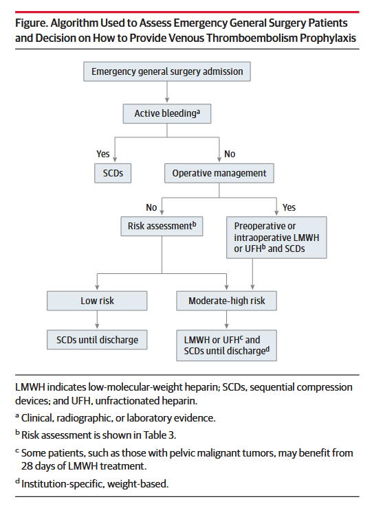
Phlegmasia alba dolens and phlegmasia cerulea dolens

Shackford SR. (2018). Venous Disease. In: Abernathy’s Surgical Secrets, 7th ed.: p. 357.

What is the difference between phlegmasia alba dolens and phlegmasia cerulea dolens? 

“These two entities occur following iliofemoral venous thrombosis, 75% of which occur on the left side presumably because of compression of the left common iliac vein by the overlying right common iliac artery (May-Thurner syndrome). Iliofemoral venous thrombosis is characterized by unilateral pain and edema of an entire lower extremity, discoloration, and groin tenderness. In phlegmasia alba dolens (literally, painful white swelling), the leg becomes pale. Arterial pulses remain normal. Progressive thrombosis may occur with propagation proximally or distally and into neighboring tributaries. The entire leg becomes both edematous and mottled or cyanotic. This stage is called phlegmasia cerulea dolens (literally, painful purple swelling). When venous outflow is seriously impeded, arterial inflow may be reduced secondarily by as much as 30%. Limb loss is a serious concern and aggressive management (i.e., venous thrombectomy, catheter-directed lytic therapy, or both) is necessary.”


Chinsakchai K, et al. Trends in management of phlegmasia cerulea dolens. Vasc Endovascular Surg. 2011 Jan;45(1):5-14. Full-text for Emory users.

PCD screenshot
Continue reading

Caprini Risk Assessment Model for DVTs

Cronin M, Dengler N, Krauss ES, et al. Completion of the Updated Caprini Risk Assessment Model (2013 Version). Clin Appl Thromb Hemost. 2019 Jan-Dec;25:1076029619838052.

Abstract: The Caprini risk assessment model (RAM) has been validated in over 250,000 patients in more than 100 clinical trials worldwide. Ultimately, appropriate treatment options are dependent on precise completion of the Caprini RAM. As the numerical score increases, the clinical venous thromboembolism rate rises exponentially in every patient group where it has been properly tested. The 2013 Caprini RAM was completed by specially trained medical students via review of the presurgical assessment history, medical clearances, and medical consults. The Caprini RAM was completed for every participant both preoperatively and predischarge to ensure that any changes in the patient’s postoperative course were captured by the tool. This process led to the development of completion guidelines to ensure consistency and accuracy of scoring. The 2013 Caprini scoring system provides a consistent, thorough, and efficacious method for risk stratification and selection of prophylaxis for the prevention of venous thrombosis.

D-dimer testing to determine the duration of anticoagulation therapy

Palareti G, Cosmi B, Legnani C, et al.; DULCIS Investigators. D-dimer to guide the duration of anticoagulation in patients with venous thromboembolism: a management study. Blood. 2014 Jul 10;124(2):196-203.

Full-text for Emory users.

The optimal duration of anticoagulation in patients with venous thromboembolism (VTE) is uncertain. We investigated whether persistently negative D-dimers in patients with vein recanalization or stable thrombotic burden can identify subjects at low recurrence risk. Outpatients with a first VTE (unprovoked or associated with weak risk factors) were eligible after at least 3 months (12 in those with residual thrombosis) of anticoagulation. They received serial D-dimer measurements using commercial assays with predefined age/sex-specific cutoffs and were followed for up to 2 years. Of 1010 patients, anticoagulation was stopped in 528 (52.3%) with persistently negative D-dimer who subsequently experienced 25 recurrences (3.0% pt-y; 95% confidence interval [CI], 2.0-4.4%). Of the remaining 482 patients, 373 resumed anticoagulation and 109 refused it. Recurrent VTE developed in 15 patients (8.8% pt-y; 95% CI, 5.0-14.1) of the latter group and in 4 of the former (0.7% pt-y; 95% CI, 0.2-1.7; hazard ratio = 2.92; 95% CI, 1.87-9.72; P = .0006). Major bleeding occurred in 14 patients (2.3% pt-y; 95% CI, 1.3-3.9) who resumed anticoagulation. Serial D-dimer measurement is suitable in clinical practice for the identification of VTE patients in whom anticoagulation can be safely discontinued. This study was registered at clinicaltrials.gov as #NCT00954395.

Continue reading

Thrombolysis for acute deep vein thrombosis

Watson L, Broderick C, Armon MP. Thrombolysis for acute deep vein thrombosis. Cochrane Database Syst Rev. 2016 Nov 10;11(11):CD002783.

Main results: Seventeen RCTs with 1103 participants were included. These studies differed in the both thrombolytic agent used and in the technique used to deliver it. Systemic, loco-regional and catheter-directed thrombolysis (CDT) were all included. Fourteen studies were rated as low risk of bias and three studies were rated as high risk of bias. We combined the results as any (all) thrombolysis compared to standard anticoagulation. Complete clot lysis occurred significantly more often in the treatment group at early follow-up (RR 4.91; 95% CI 1.66 to 14.53, P = 0.004) and at intermediate follow-up (RR 2.44; 95% CI 1.40 to 4.27, P = 0.002; moderate quality evidence). A similar effect was seen for any degree of improvement in venous patency. Up to five years after treatment significantly less PTS occurred in those receiving thrombolysis (RR 0.66, 95% CI 0.53 to 0.81; P < 0.0001; moderate quality evidence). This reduction in PTS was still observed at late follow-up (beyond five years), in two studies (RR 0.58, 95% CI 0.45 to 0.77; P < 0.0001; moderate quality evidence). Leg ulceration was reduced although the data were limited by small numbers (RR 0.87; 95% CI 0.16 to 4.73, P = 0.87). Those receiving thrombolysis had increased bleeding complications (RR 2.23; 95% CI 1.41 to 3.52, P = 0.0006; moderate quality evidence). Three strokes occurred in the treatment group, all in trials conducted pre-1990, and none in the control group. There was no significant effect on mortality detected at either early or intermediate follow-up. Data on the occurrence of pulmonary embolism (PE) and recurrent DVT were inconclusive. Systemic thrombolysis and CDT had similar levels of effectiveness. Studies of CDT included two trials in femoral and iliofemoral DVT, and results from these are consistent with those from trials of systemic thrombolysis in DVT at other levels of occlusion.

Continue reading

Extended-duration thromboprophylaxis after CRS/HIPEC

Khan S, et al. Incidence, Risk Factors, and Prevention Strategies for Venous Thromboembolism after Cytoreductive Surgery and Hyperthermic Intraperitoneal Chemotherapy. Ann Surg Oncol. 2019 Jul;26(7):2276-2284.

Full-text for Emory users.

“A policy change was made in February 2010 to discharge all patients post-CRS/HIPEC with 14 days of additional pharmacothromboprophylaxis, which consisted of low-molecular-weight heparin in 327 of 447 (73%) cases (Supplemental Figure). The 60-day VTE rate decreased from 10.2 to 4.9% after this policy was instituted (p = 0.10, Fig. 2).”

“This policy is in accordance with established guidelines indicating the need for a total of 4 weeks of pharmacothromboprophylaxis in high-risk patients after abdominal or pelvic surgery for cancer. [2,21] Given that patients have an average length of stay of nearly 2 weeks, discharging them on 14 days of pharmacothromboprophylaxis fulfills this duration.”

Continue reading

CME & Education: VTE Prevention in the Hospital: New Approaches and Expert Perspectives

This continuing education offering is part of Medscape‘s series, Contemporary Topics in Antithrombotic Therapy. (You’ll need a Medscape account to view and/or accrue CME credit.)

Authors: Gary E. Raskob, PhD; Steven B. Deitelzweig, MD; Alex C. Spyropoulos, MD

CME Released: 12/22/2019; Valid for credit through: 12/22/2020

“…[W]e are going to talk about VTE, its importance in the hospital population of patients admitted with medical illness, and how we can work to reduce the burden of disease from this important condition.

About half of all hospitalizations in the United States are for medical illnesses, such as heart failure, pneumonia, stroke, and so on. Of these patients, about half of them are at risk for VTE and about 25% are at high risk for VTE.

Those who develop VTE tend to have pretty severe consequences, and these consequences persist beyond hospitalization.”