Article of interest: Volvulus of the Small Bowel and Colon

Kapadia MR. Volvulus of the Small Bowel and Colon. Clin Colon Rectal Surg. 2017 Feb; 30(1):40-45.

Free full-text.

“Volvulus of the intestines involves twisting around a fixed point. It may occur anywhere along the gastrointestinal tract where there is a long, mobile intestinal segment with a narrow mesenteric attachment. Volvulus leads to luminal obstruction and can compromise intestinal blood flow. For this reason, it tends to be a surgical emergency which requires prompt attention. Failure to recognize the signs and symptoms of intestinal volvulus may lead to bowel ischemia and perforation.”

Continue reading →

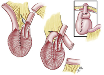
Continent ileostomy modifications

Today’s discussion topics included the Barnett continent intestinal reservoir.


Worsey J, Fazio VW. (2017). Continent Ileostomy. In: Current Therapy in Colon and Rectal Surgery, 3rd ed, pgs. 204-208. Elsevier: NY.

Full-text for Emory users.

Philadelphia Illustration Dept./Elsevier

FIGURE 40-3. Barnett modification of the continent ileostomy. (Cleveland Clinic Center for Medical Art & Photography.)

Continue reading →

Stoma versus stent as a bridge to surgery for obstructive colon cancer

Veld JV, et al. Changes in Management of Left-Sided Obstructive Colon Cancer: National Practice and Guideline Implementation. J Natl Compr Canc Netw. 2019 Dec;17(12):1512-1520.

Free full-text. 

Results: A total of 2,587 patients were included (2,013 ER, 345 DS, and 229 SEMS). A trend was observed in reversal of ER (decrease from 86.2% to 69.6%) and SEMS (increase from 1.3% to 7.8%) after 2014, with an ongoing increase in DS (from 5.2% in 2009 to 22.7% in 2016). DS after 2014 was associated with more laparoscopic resections (66.0% vs 35.5%; P<.001) and more 2-stage procedures (41.5% vs 28.6%; P=.01) with fewer permanent stomas (14.7% vs 29.5%; P=.005). Overall, more laparoscopic resections (25.4% vs 13.2%; P<.001) and shorter total hospital stays (14 vs 15 days; P<.001) were observed after 2014. However, similar rates of primary anastomosis (48.7% vs 48.6%; P=.961), 90-day complications (40.4% vs 37.9%; P=.254), and 90-day mortality (6.5% vs 7.0%; P=.635) were observed.

CONCLUSIONS: Guideline revision resulted in a notable change from ER to BTS for LSOCC. This was accompanied by an increased rate of laparoscopic resections, more 2-stage procedures with a decreased permanent stoma rate in patients receiving DS as BTS, and a shorter total hospital stay. However, overall 90-day complication and mortality rates remained relatively high.

Continue reading →

Pathogenesis of diversion colitis

This week’s discussions included the causes of diversion colitis.


Tominaga K, et al. Diversion colitis and pouchitis: A mini-review. World J Gastroenterol. 2018 Apr 28;24(16):1734-1747.

Free full-text.

“The basic mechanisms underlying diversion colitis are still unclear. Glotzer hypothesized that it might be the result of bacterial overgrowth, the presence of harmful bacteria, nutritional deficiencies, toxins, or disturbance in the symbiotic relationship between luminal bacteria and the mucosal layer[2]. Reportedly, concentrations of carbohydrate-fermenting anaerobic bacteria and pathogenic bacteria are reduced in de-functioned colons[5,23,53] and these reports indicate that the overgrowth of anaerobic bacteria or a pathogenic bacterium is unlikely to be an important etiological factor. On the other hand, there is an increase of nitrate-reducing bacteria in patients with diversion colitis[7] and nitrate-reducing bacteria produce nitric oxide (NO) which plays a protective role in low concentrations, but at higher levels it becomes toxic to the colonic tissue[54]. Thus, it has been suggested that increases in nitrate-reducing bacteria may result in toxic levels of NO, leading to the diversion colitis.” (Tominaga, 2018, p. 1739)

Continue reading →

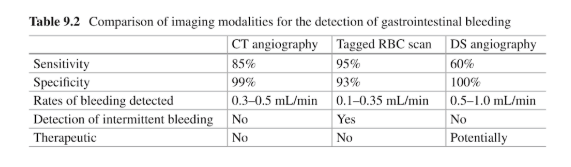
Upper GI bleeding: CTA prior to flouroscopic angiography?

A discussion this week included a diagnostic CTA prior to flourscopic angiography.


Reference: Wells ML, et al. CT for evaluation of acute gastrointestinal bleeding. RadioGraphics. 2018 Jul-Aug;38(4):1089-1107. doi:10.1148/rg.2018170138

Summary: “Teaching point: CT angiography is gaining popularity for use in emergent evaluations of acute GI bleeding. It has potential for use in the first-line evaluation of acute LGIB and the evaluation of UGIB after failed or nondiagnostic endoscopy.”

Continue reading →

Are diabetic patients at greater risk for anastomotic leaks and mortality when undergoing colectomies?

One discussion this week included postoperative anastomotic leaks.

Reference: Ziegler MA, et al. Risk factors for anastomotic leak and mortality in diabetic patients undergoing colectomy: analysis from a statewide surgical quality collaborative. Archives of Surgery. 2012 Jul;147(7):600-605. doi: 10.1001/archsurg.2012.77.

Summary: In a database review of patients in Michigan who underwent colectomy, the study aimed to determine risk factors in diabetic patients that are associated with increased postcolectomy mortality and anastomotic leak.

Primary risk factors were diabetes mellitus, hyperglycemia, steroid use, and emergency surgery. Of the 5123 patients, 889 were diabetic, 4234 were nondiabetic.

Diabetes alone was not found to be a risk factor for anastomotic leak in this study.

  • 56% of diabetic patients had preoperative glucose levels of 140 mg/dL or higher
  • Preoperative steroid use led to increased rates of anastomotic leak in diabetic patients
  • Diabetic patients who had a leak had more than a 4-fold higher mortality (26.3% vs 4.5%, P<.001) compared with nondiabetic patients (6.0% vs 2.5%, P<.05).
  • Mortality was associated with hyperglycemia for nondiabetic patients only

The authors conclude that improved screening may identify high-risk patients who would benefit from perioperative intervention.