High Prevalence and Mortality Associated with Upper Extremity Deep Venous Thrombosis in Hospitalized Patients at a Tertiary Care Center

“Venous thromboembolism (VTE), which includes deep venous thrombosis (DVT) and pulmonary embolism (PE), is the third most common cardiovascular disorder with an annual incidence of 0.1%, affecting approximately 5% of the population.1 Upper extremity DVT (UEDVT) has been thought to account for 4e10% of all cases of DVT and may involve the radial, ulnar, brachial, axillary, subclavian, brachiocephalic, or internal jugular veins.
Historically, UEDVT has been considered a relatively benign event. However, as the incidence of UEDVT increases, so do its subsequent complications including PE, venous access difficulties, superior vena cava syndrome, postthrombotic syndrome, and bleeding on therapeutic anticoagulation therapy, suggesting an insufficient understanding and management of UEDVT.
Management guidelines, with the exception of those for thoracic outlet syndrome, are largely
extrapolated from lower extremity DVT (LEDVT) and PE management.”

Continue reading

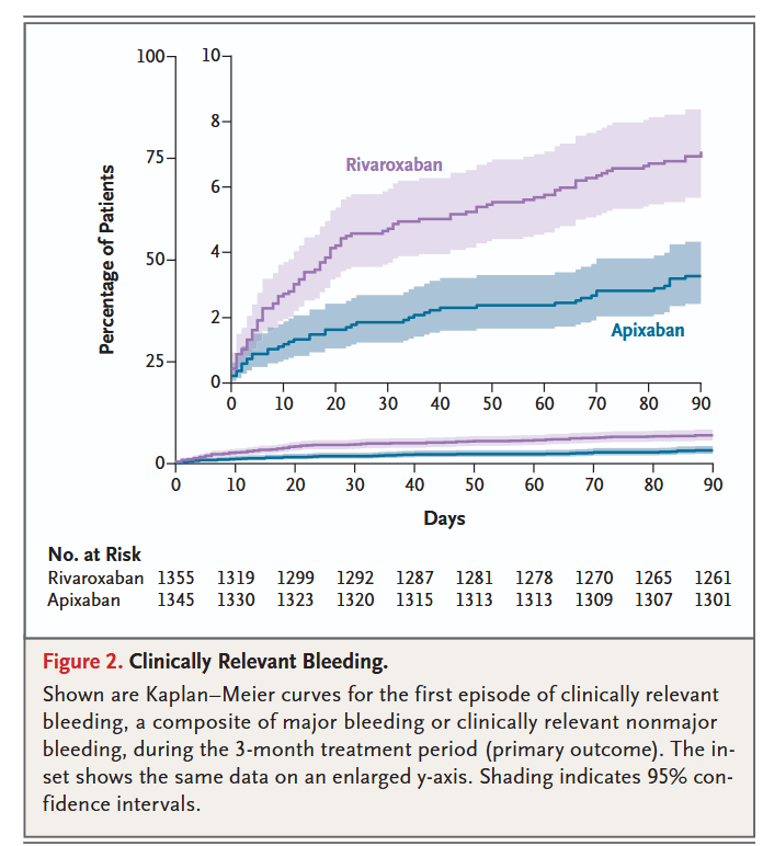
Anticoagulant treatment for upper extremity deep vein thrombosis

“Upper extremity DVT may be complicated by recurrent thrombosis in about 7.5% of cases and by post-thrombotic syndrome in 19.4% of cases, with higher rates of recurrence in patients with cancer-associated thrombosis and of post-thrombotic syndrome in patients with unprovoked thrombosis or related to congenital or anatomical abnormalities. The intensity and duration of anticoagulant treatment need to be balanced against the risk of bleeding
complications, especially in high-risk subgroups like patients with cancer in whom major bleeding events were reported in up to 10%.”

FIGURE 2 Recurrent venous thromboembolism and major bleeding in patients with upper extremity deep vein thrombosis, sorted by the proportion of patients with cancer and an indwelling catheter. Recurrent venous thromboembolism and major bleeding occurring during anticoagulant treatment were considered in the analysis. The vertical line indicates the summary estimate. Gray squares indicate individual study estimates of the proportion, whereas the gray horizontal lines indicate 95% confidence intervals of the individual studies. The diamond indicates the summary estimate with 95% confidence intervals. The horizontal black line refers to the prediction intervals which are displayed numerically under the 95% confidence intervals. CI, confidence interval; ES, estimates; PI, prediction interval
Continue reading

Routine sterile glove and instrument change at the time of abdominal wound closure to prevent surgical site infection (ChEETAh)

“Surgical site infection (SSI) is the most common complication of surgery around the world, and disproportionately affects patients in low income and middle income countries (LMICs). SSI is unpleasant and harmful for patients, increases the care burden on families and communities, and is very costly for patients and providers. As a result, SSI was highlighted as the highest research priority in surgery in a global co prioritisation exercise. The causes of SSI are multifactorial, and a so called magic bullet to prevent SSI is unlikely to exist. In modern surgical practice, very few interventions to reduce the incidence of SSI have
been shown to be effective when tested robustly.”

“The ChEETAh trial evaluated a change in behaviour across theatre teams to routinely change gloves and instruments at the time of abdominal wound closure. Compared with
previous studies, ChEETAh is large, pragmatic, rigorously conducted, and transparently reported. It also includes a diverse and representative range of patients operated on in
hospitals across middle-income and lower-income settings. The high adherence rate with the intervention shows that routine change of gloves and instruments is deliverable
around the world. ChEETah robustly shows that routine change of gloves and instruments before wound closure reduced surgical site infections in clean–contaminated, contaminated, and dirty surgery, which was consistent across several sensitivity analyses. That there was no evidence to suggest heterogeneity of effect across any of the prespecified subgroups suggests that the effect is consistent across a wide range of patients.”

Continue reading

Symptomatic perioperative venous thromboembolism is a frequent complication inpatients with a history of deep vein thrombosis

“Perioperative venous thromboembolism (VTE) is a frequent complication in patients who undergo major surgical procedures. The risk for thromboembolism is increased in patients with advancing age, malignancy, inherited and acquired thrombophilic conditions, and in those patients who undergo certain higher-risk surgical procedures (for example: total hip arthroplasty, total knee arthroplasty, and abdominal cancer operations). A history of deep vein thrombosis (DVT) or pulmonary embolism (PE) frequently is cited as a risk factor for subsequent recurrence around the time of surgery. However, the incidence of perioperative VTE recurrence in patients with a history of lower extremity deep vein thrombosis is not well studied.”

Continue reading

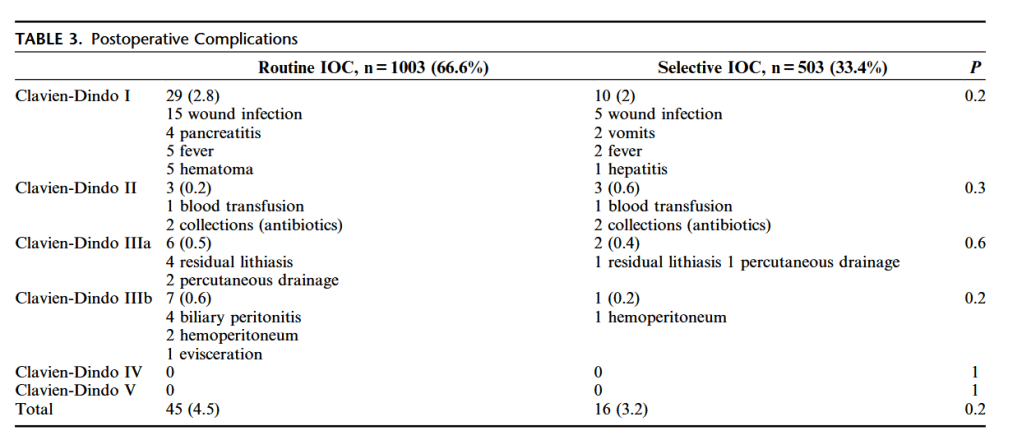
Operative Outcomes for Polycystic Liver Disease

“Currently, there are no effective medical therapies proven to benefit symptomatic PCLD. Some data exist to support the use of somatostatin analogs in the treatment of hepatomegaly due to PCLD; however, the durability of their efficacy remain in question. Vasopressin receptor antagonists, ursodeoxycholic acid, and protein kinase inhibitors
have also been investigated in the management of symptomatic PCLD, but none has been shown to reduce liver volume sufficiently for symptom improvement. With the lack of medical therapies, management of advanced PCLD relies on procedural intervention. Cyst aspiration may be sufficient in patients with a single large cyst; however, with the innumerable cysts present in PCLD, aspiration is frequently ineffective.”

Continue reading

Laparoscopic vs. open feeding jejunostomy

“Feeding jejunostomy is conventionally done via laparotomy. However, since laparoscopic jejunostomy was introduced in 1990, the procedure is constantly advancing with new techniques and devices. It was demonstrated to be a safe, feasible, and cost-effective technique with comparable complication rate to open feeding jejunostomy.”

“In addition, compared with the conventional open procedure, the laparoscopic approach has the inherit merits of smaller incisions, better cosmesis, less postoperative pain, and earlier recovery. With the aim of achieving early enteral feeding and a reduction in postoperative morbidity, any complications arising from the procedure will jeopardize its benefits, incur additional costs, and delay subsequent oncologic treatment.”

Continue reading

Safety of mechanical chest compression devices AutoPulse and LUCAS in cardiac arrest

“Mechanical chest compression devices are designed to perform chest compressions at specified rate and depth and therefore were expected to improve outcome. There are at
present two widely used and Food and Drug Administration-approved devices: the AutoPulse, a load-distributed band device that rhythmically compresses and restricts the chest wall and the LUCAS, a piston device with a cup that is placed in the centre of the chest and pushes the sternum down over a distance of 5.2 cm and pulls back to the neutral position. Significant improvement of aortic blood pressure and coronary perfusion pressure is documented in humans from the AutoPulse compared with manual chest compressions. Chest compression with LUCAS resulted in significantly higher end-tidal carbon dioxide in humans compared with manual chest compressions. For several years, only one randomized clinical trial with the AutoPulse was available (ASPIRE), which was terminated after interim analysis because of a trend to reduced survival to discharge compared with manual control CPR. None of the more recent randomized clinical trials demonstrated survival benefit of AutoPulse or LUCAS over manual controls. Anecdotal and possibly biased observations in our hospital and a published letter suggested increased
damage caused by mechanical chest compression devices.”

Continue reading